Venous Ulcers can look deceptively small: a shallow break near the ankle, surrounded by itchy, swollen skin. Yet healing may take months, and recurrence is common when the underlying venous pressure remains untreated. In 2026, the best approach is not one dressing or device. It is a plan that addresses circulation, protects the wound, and fits the person’s health and daily life.
Wound-care expert Dr. Robert Kirsner is associated with a useful cause-focused principle, paraphrased here: “Treat the cause, not only the wound.” Compression is often central to care, but it should follow clinical assessment, including checks for arterial disease. A clinician may also recommend dressings, leg elevation, movement, and treatment of superficial venous reflux. The right combination depends on the ulcer and the patient. Details matter.
This article examines what current evidence can—and cannot—say about the best treatment for Venous Ulcers. It compares compression options, wound-care practices, and procedures that may reduce recurrence. It also explains when specialist assessment is important. Healing is rarely a straight line. That can be frustrating. Even careful plans may need adjustment when pain, swelling, skin changes, or slow progress appear. The aim is practical guidance, not a promise of quick healing: readers should leave better prepared to discuss safe, individualized care with a qualified clinician.
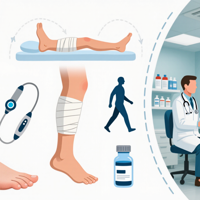
Venous ulcers are not diagnosed by appearance alone. A shallow, irregular wound near the inner ankle, swelling, visible veins, or brown, itchy skin can suggest chronic venous disease. But arterial disease, diabetes, pressure injury, and infection can look similar or occur alongside it.
The European Society for Vascular Surgery’s 2022 guidelines estimate that venous disease causes about 70% of leg ulcers. That makes careful assessment essential.
Clinicians review medical history and record the wound’s location, size, depth, drainage, and surrounding skin.
Look beyond the wound.
Assessment may include checking pulses and measuring the ankle-brachial index (ABI), which compares ankle and arm blood pressure. This helps identify reduced arterial circulation before compression is considered.
Duplex ultrasound can assess venous reflux or obstruction; the ESVS guidelines identify it as the primary imaging test for chronic venous disease. Pain, swelling, and changes in skin temperature also matter.
One limitation: findings do not always point to a single cause, so reassessment may be needed as the wound changes.
Tips: Note when swelling worsens and what eases it. Bring a current medication list and any wound measurements to appointments. Seek prompt clinical advice for spreading redness, fever, or sudden increases in pain.
For venous leg ulcers in 2026, compression is usually the treatment foundation, not an optional extra. It helps counter the raised vein pressure that drives swelling and delays healing. A 2021 Cochrane review examined 14 randomized trials involving 1,391 people. Compression probably increased the chance of complete healing within 12 months compared with no compression (risk ratio 2.17; 95% CI 1.52–3.10). That is meaningful evidence, though it does not guarantee a wound will close. Small details matter.
The right pressure depends on the person, not just the ulcer. Clinicians should assess leg circulation before applying firm compression, since significant arterial disease can make it unsafe. Bandages or fitted stockings need to suit the wound, swelling, skin condition, and ability to use them consistently. A poorly fitted wrap can slip, bunch behind the ankle, or cause pressure pain. Not a cure-all. Movement, leg elevation, and suitable wound dressings may support care, but they do not replace compression when it is appropriate. New numbness, increasing pain, or pale, cool toes warrant prompt clinical review. Even good plans need adjustment as swelling changes.
| Treatment dimension | Evidence-based approach | Practical details | Key safety or follow-up point |
|---|---|---|---|
| Confirm the cause and assess circulation | Assess the ulcer and surrounding skin, check leg pulses, and evaluate for venous disease. Measure the ankle–brachial pressure index (ABPI) or use another appropriate arterial assessment before applying strong compression. | Consider duplex ultrasound when venous reflux or obstruction needs assessment, or when planning a venous procedure. Investigate atypical, rapidly worsening, or non-healing ulcers for other causes. | Compression strength must be matched to arterial circulation and the person’s overall clinical condition. ABPI interpretation can be unreliable in some people, including those with heavily calcified arteries. |
| Compression therapy Foundation of treatment | For a confirmed venous ulcer with adequate arterial supply, use therapeutic compression to reduce venous hypertension and swelling. Multilayer bandaging or a suitable compression system can be selected according to the patient and care setting. | When clinically appropriate, commonly used high-compression systems aim for about 30–40 mmHg at the ankle. A trained clinician should select, apply, and monitor the system; fit and consistent use matter. | Do not apply routine high compression without checking arterial supply. Severe peripheral arterial disease, significant heart failure, neuropathy, or intolerance requires individualized assessment. Stop and seek clinical advice for new severe pain, numbness, colour change, or a cold foot. |
| Modified compression when arterial disease is present | Some people with mixed venous and arterial disease may be considered for reduced or modified compression under specialist supervision. | Thresholds vary across guidelines and clinical circumstances. An ABPI around 0.5–0.8 may prompt consideration of modified compression with close monitoring; absolute ankle and toe pressures and the overall assessment also matter. | With very low arterial pressures—for example, ABPI below about 0.5, ankle pressure below 60 mmHg, or toe pressure below 30 mmHg—sustained compression is generally avoided pending vascular assessment. |
| Wound cleansing and dressings | Cleanse the wound and use an appropriate dressing to manage exudate and protect the wound and surrounding skin. Compression, rather than a particular dressing type, is the key treatment for venous hypertension. | Choose a dressing based on wound moisture, comfort, skin condition, and how often the compression system needs changing. Protect fragile or macerated surrounding skin. | No single dressing type has been shown to be best for every venous ulcer. Reassess if the wound deteriorates, exudate changes substantially, or contact sensitivity is suspected. |
| Leg elevation and mobility | Encourage regular walking and ankle movement when feasible, alongside compression. Elevating the leg while resting can help reduce dependent swelling. | Adapt activity and elevation advice to mobility, balance, pain, and other health conditions. | These measures support care but do not replace appropriate compression or assessment of the underlying venous disease. |
| Infection management | Use antibiotics only when there are clinical signs of infection, such as spreading redness or warmth, increasing pain, fever, or systemic illness. | Assess the patient and wound clinically; select treatment according to local guidance and the likely infection. | Colonization or a positive wound culture alone does not necessarily mean antibiotics are needed. Urgent assessment is appropriate for systemic illness or rapidly spreading infection. |
| Venous intervention | For suitable patients with superficial venous reflux, early endovenous treatment alongside compression can accelerate healing compared with compression while waiting for intervention. | Duplex ultrasound helps identify reflux and guide assessment for procedures such as endovenous ablation. Suitability depends on anatomy, health, and patient preference. | Intervention complements rather than automatically replaces wound care and compression. Discuss benefits, risks, and alternatives with a vascular specialist. |
| Adjunctive medicines | Pentoxifylline may be considered as an adjunct to compression for selected patients, subject to local guidance and individual contraindications. | Review medication interactions, side effects, and the patient’s clinical history before use. | Medicines do not replace compression when compression is safe and appropriate. Routine antibiotics are not a treatment for an uninfected ulcer. |
| Review, healing, and recurrence prevention | Monitor wound size, pain, swelling, skin condition, and tolerance of compression. After healing, ongoing compression hosiery is often recommended to reduce recurrence when arterial assessment and fit allow. | Review progress regularly and check the fit and condition of bandages or hosiery. Reassess the diagnosis and care plan if the ulcer fails to improve or worsens. | Recurrence prevention usually requires continued attention to venous disease, skin care, mobility, and long-term compression where suitable. |
| Clinical note: This table is a general educational overview, not an individualized treatment plan. Compression pressure, arterial thresholds, and treatment choices should be determined by a qualified clinician using current local guidance and a complete assessment. | |||
Venous ulcers often appear near the ankle, where swelling and poor blood flow can leave skin fragile. Treatment usually combines compression with regular wound care. Compression can reduce swelling and support blood return, but a clinician should check leg circulation before recommending it. The right plan depends on the ulcer, overall health, and any other circulation problems.
Clean, careful wound care helps protect the ulcer as new tissue forms. A clinician may rinse it with a suitable solution, cover it with a dressing that manages drainage, and change that dressing on a set schedule. Avoid scrubbing or applying unprescribed creams. Small details count. A damp, leaking bandage or newly irritated skin deserves attention, not guesswork.
At home, follow the care plan, elevate the leg when resting, and take gentle walks if safe for you. Healing can be slow, and progress is not always neat or steady. That can be frustrating. Keep follow-up visits so the wound and compression can be reassessed. Increasing redness, warmth, pain, or drainage may signal infection; fever or rapidly worsening symptoms need prompt medical attention. Antibiotics are generally used when a clinician finds an infection, not simply because an ulcer is present.
Advanced treatment is considered when an ulcer remains open despite consistent compression, wound care, and attention to mobility. A procedure is not always the next step. Clinicians check circulation and may measure ankle-brachial pressure before recommending stronger compression, because arterial disease can coexist. A duplex ultrasound can reveal whether faulty vein valves are sending blood backward. That detail matters.
When superficial venous reflux is confirmed, endovenous ablation may be an option. It closes the affected vein and, for suitable patients, can support faster healing when paired with compression. It does not replace ongoing compression. Results are not guaranteed, and some people are not candidates. Ask about possible pain, skin changes, clot risk, and recovery time.
For a wound that still will not progress, a wound specialist may consider debridement, a skin graft, or a tissue-based treatment. These options are not interchangeable, and evidence varies by ulcer type. Antibiotics are appropriate when clinical signs indicate infection; they do not heal an uninfected ulcer. Keep track of wound size, drainage, and pain, and ask what changes would show the plan is working. Sometimes the plan needs revision.
Preventing a venous ulcer from returning often requires ongoing compression, even after the skin closes.
A clinician should check circulation before compression begins, since poor arterial blood flow can make it unsafe. The right garment or bandage depends on the person’s leg shape, comfort, and ability to use it. A snug fit matters. If it slips, bunches, or causes new pain, ask for a review rather than simply tightening it. Daily walking and ankle movements can support the calf-muscle pump; raising the legs when resting may also ease swelling. Small changes matter.
Long-term monitoring should look beyond the wound itself. At follow-up visits, a care team can measure the area, note drainage, and compare dated photographs with consent. At home, watch for renewed swelling, skin darkening, itching, or a tender spot that does not improve. Seek prompt medical advice for spreading redness, increasing pain, fever, or sudden deterioration. Healing is rarely linear. A missed day of compression does not always mean failure, but repeated difficulties deserve a practical plan. Some people struggle with heat, dexterity, or finding a comfortable fit; that should be discussed honestly. Review the plan regularly, especially if swelling or mobility changes, and keep follow-up appointments even when the wound looks closed.